RADIO VISION – SCIENTIFIC MILESTONE
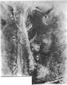
The Sacral Plexus Region
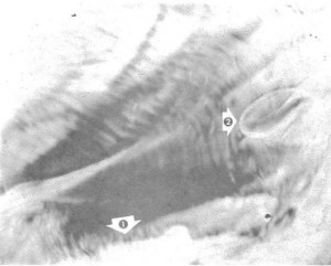
Arrow 1 shows a cross section of the sacral bone. Arrow 2 shows portions of the vertebrae. It is as though the patient were facing the left side of the picture, and his pelvic region vertically cross sectioned by a gigantic knife. Arrow 3 shows three blood vessels in a state of diapedesis, carrying blood to the meningeal coccus infection concentrated at the ends of the blood vessels near the vertebrae. The meningeal coccus, on which the Radio-Vision Instrument was tuned, shows as myriad black dots all through the region photographed. The infection shows in the bottom right hand corner, in the sacral bone itself; it shows through the structure of the gluteal muscle (Arrow 4). The black circles, (Arrow 5) are blood vessels which travel at right angles to the plane of the picture.
The infection was detected first by the Drown Diagnostic Instrument. Then the Radio-Vision Instrument was tuned in on the infection, using the rate established in the diagnostic process. The patient was seven city blocks away from the instrument when the picture was made.
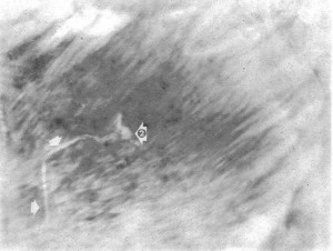
Gallstone in Gall Bladder.
This Radio-Vision photograph was made by tuning in on the gallstone, detected in the Drown diagnosis. The photograph is a perfect cross section of the gallbladder, the stone itself appearing imbedded in the tissue (Arrow 1) The inflammation (arrow 2) shows above the stone itself. The almost three dimensional nature of the picture shows the startling clarity with which these soft tissue pictures may be made. The patient was separated from the instrument by one floor of a building and connected to it only by her blood crystal.
This Radio-Vision photograph shows the Thyroid Gland (arrow A) in a case of Hodgkins disease. The two arrows (2) show the spinal cord, deleting the vertebral bony structure. The numerous black circles indicated by arrow 3 are cross sections of blood vessels in the pleura of the lungs. Arrow 4 indicates the pleura.
The patient was 1300 miles away when the picture was made via the blood crystal, Drown diagnosis having previously verified the presence of Hodgkins’ disease.
Pathway of Bullet in Patient’s Chest
A domestic servant of Dr. Drown in the years before World War II feared that her son had been injured by a rifle shot, aimed at him indiscriminately by young hoodlums passing in a car. Knowing of Dr. Drown’s photographic instrument, she brought a crystal of the boy’s blood to Dr. Drown.
Dr. Drown took two photographs by Radio-Vision. The first photograph, this one, saw the Radio-Vision Instrument set to the injury or wound rate. The arrows indicate the pathway through the tissue of the chest followed by the bullet, which had in fact lodged in the boy’s body. Arrow 2 indicates the area of final lodgement of the projectile.
Actual Bullet in Patient’s Chest
This Radio-Vision photograph was made immediately following plate 4. The tuning of the Radio-Vision Instrument was altered to tune in directly on the bullet itself, rather than on the wound. The arrow shows the actual bullet end-on, and it measures on the original 8 X 10 photograph exactly .22 inches.
The boy with the bullet in his chest was 22 miles away at the time this Radio-Vision photograph was made, the only connection with the Radio-Vision Instrument being his blood crystal, supplied by his mother.
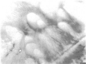
Ovum in an Ovary
This Radio-Vision photograph shows Oophorons in different phases of development in an ovary. The vivid, egg— shapes are undeniable. Arrow shows a ruptured oophoron in the lower left corner of the picture.
Cancer of the Stomach
This picture was thought by a number of doctors to be a diaphragmatic hemia. However, the Drown diagnosis recorded cancer of the area shown, and it was into this that the Radio-Vision Instrument was tuned. The arrows show the outline of the cancer. Numerous blood vessels show as black spots and spheres, as they run at right angles to the plane of the photograph.
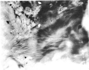
Ulcer of the Stomach and Scirrhous Cancer
This Radio-Vision photograph is a cross sectional picture of the stomach of a patient whose diagnosis showed both scirrhous cancer and an ulcer. The cancer shows as the white, thickened tissue (Arrow 1), the ulcer on the right being clearly outlined (Arrow 2). Photograph made from patient’s blood crystal, with patient remote geographically from the Radio-Vision Instrument.


